Understanding the condition
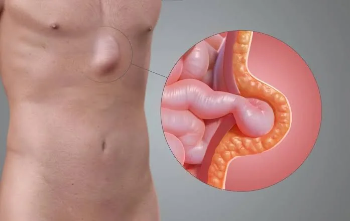
An umbilical hernia forms at or near the navel. An epigastric hernia forms in the midline between the navel and breastbone. Fat often protrudes through a small gap; larger hernias may contain bowel.
A widening between the abdominal muscles, called rectus diastasis, can occur alongside a hernia. It is not itself a true hernia, but it can affect repair planning and recurrence risk.
How it is assessed
Examination usually confirms the diagnosis and measures the defect. Ultrasound can clarify a small or uncertain lump. CT is useful for larger, recurrent or multiple defects. The surgeon also reviews skin condition, body mass index, diabetes, smoking, previous surgery and pregnancy plans.
When is surgery considered?
Repair is commonly advised when the hernia is painful, enlarging, difficult to reduce or limiting activity. In adults it will not close by itself and tends to enlarge, so repair is usually advised.
Smoking cessation, diabetes control and an achievable weight plan can lower wound risk before elective surgery. Timing around a future pregnancy should also be discussed because pregnancy can stretch a repair.
Surgery and important risks
Repair returns the contents to the abdomen and closes the defect. Sutures alone may suit very small defects in selected patients. For defects around 1 cm or larger, a mesh is usually used to reinforce the repair. Repair may be open, laparoscopic or robotic. Options include IPOM Plus, Ventral TAPP (laparoscopic or robotic), PeTEP and sublay (retrorectus) repair. The choice depends on the size of the gap, separated abdominal muscles (divarication) and any previous surgery.
Risks include seroma, haematoma, wound infection, skin or navel complications, bowel injury, persistent discomfort and recurrence. The surgeon explains where the mesh will sit to best support your abdominal wall.
Recovery and follow-up
The length of stay depends on the size of the repair. Early walking and deep breathing are encouraged. Support the wound when coughing and avoid constipation. Return to work and lifting depends on pain, wound healing and physical demands.
Contact the team for fever, spreading redness, wound discharge, vomiting, worsening pain or a swelling that becomes hard. A soft postoperative fluid collection may settle, but it should not be punctured at home.
When to Seek Urgent Care
Seek emergency care if the lump becomes suddenly painful, tense or impossible to push back, especially with vomiting, abdominal swelling, redness, fever or inability to pass stool or gas.